Understanding hypertension-related brain haemorrhage through comics
Hypertension is a familiar term.
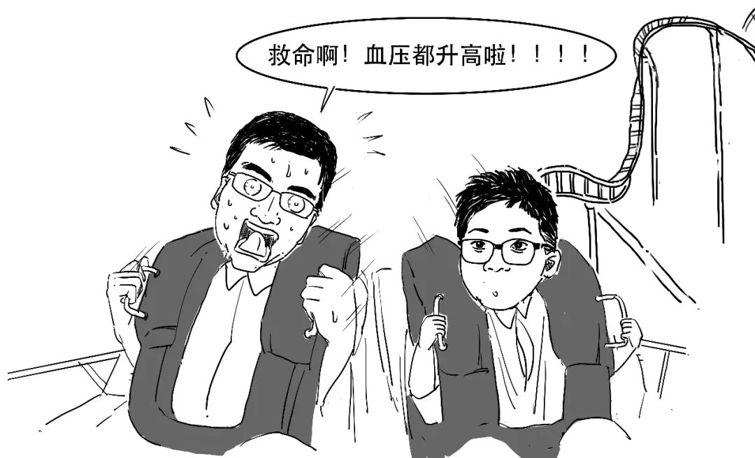
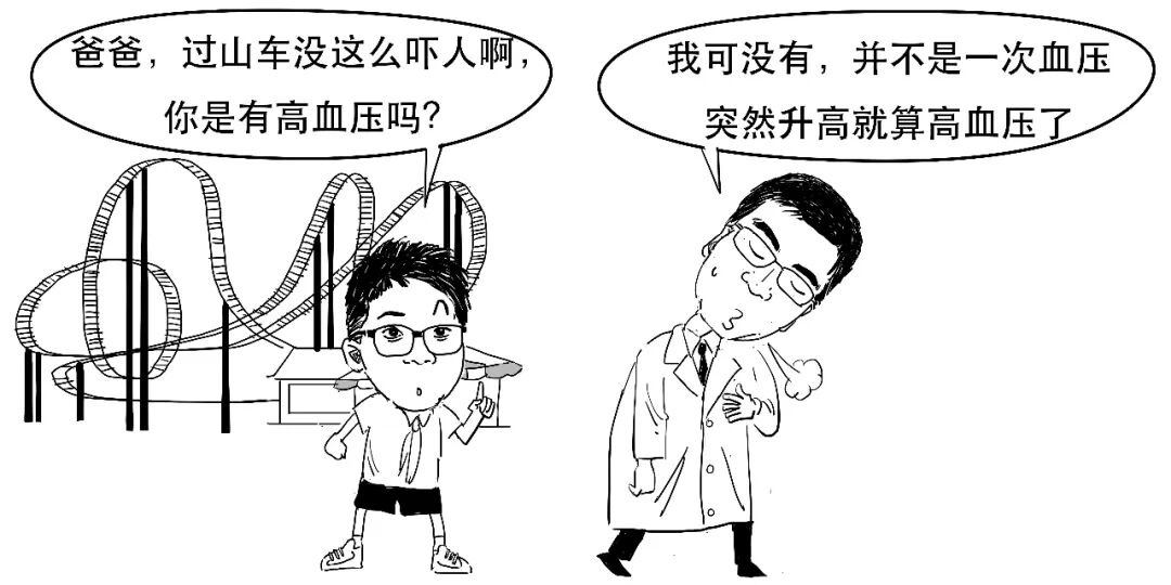
The article explains that identifying hypertension requires two measurements taken at separate times, both showing high blood pressure. Today we discuss hypertension and the brain haemorrhage it can cause.
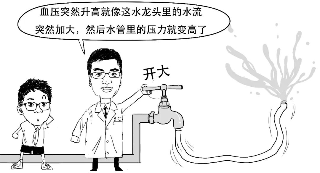
Hypertension is a long-term condition, like a tap continually turned up high and maintaining high pressure in a pipe.
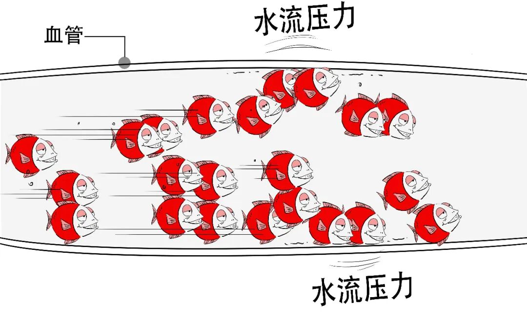
Blood flows through vessels like water through pipes and exerts pressure on their walls.
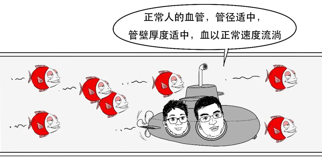
In people with hypertension, altered blood flow exerts greater pressure, gradually weakening some parts of the vessels.
How high is “high”? Blood pressure includes systolic and diastolic pressure: the pressure on vessel walls when the heart contracts and when it relaxes.
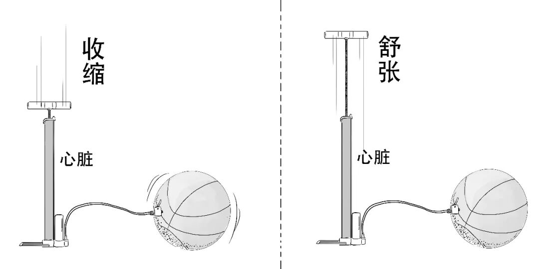
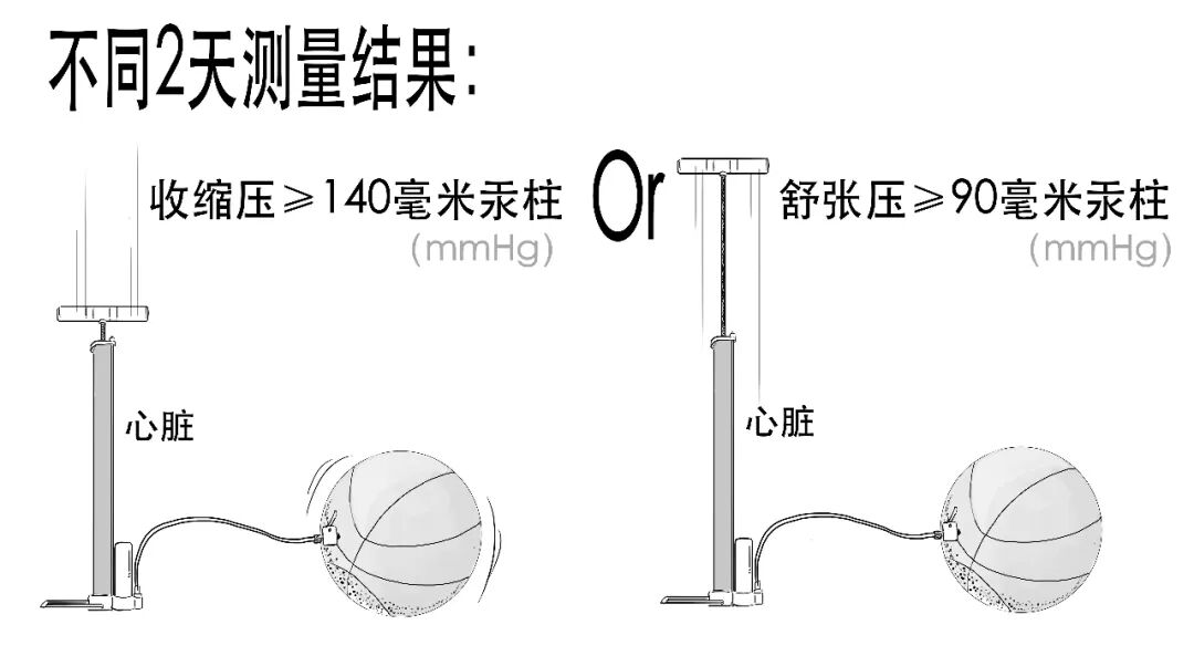
Like inflating a balloon, systolic pressure is greater than diastolic pressure. Hypertension means systolic or diastolic pressure reaches a particular level, as illustrated below.
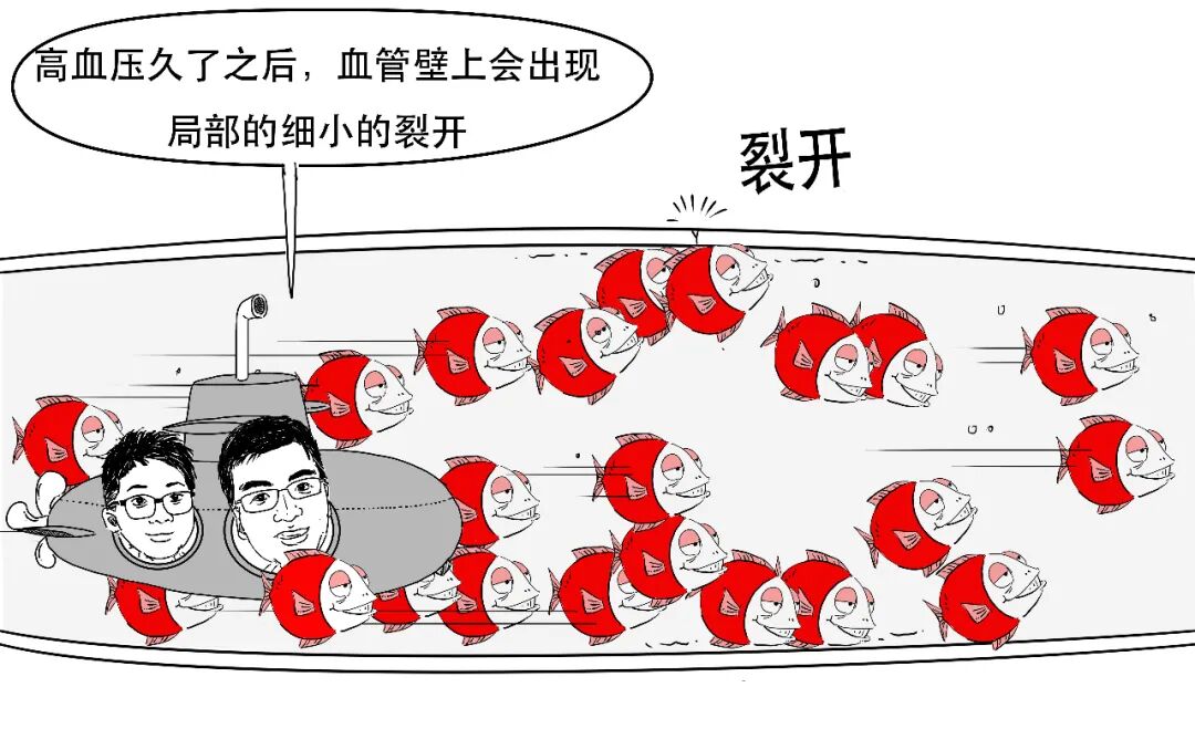
If vessel walls have been damaged by sustained pressure, a sudden rise may cause rupture and bleeding.
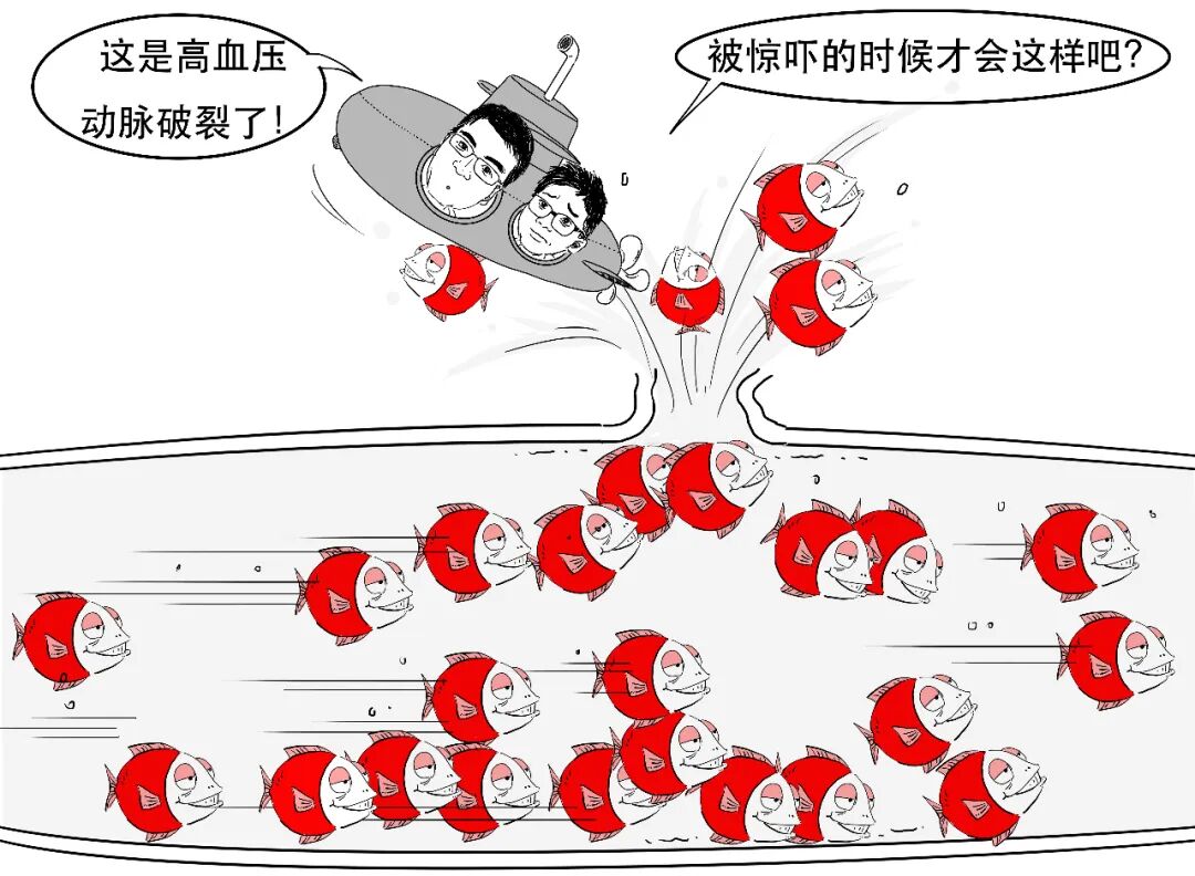
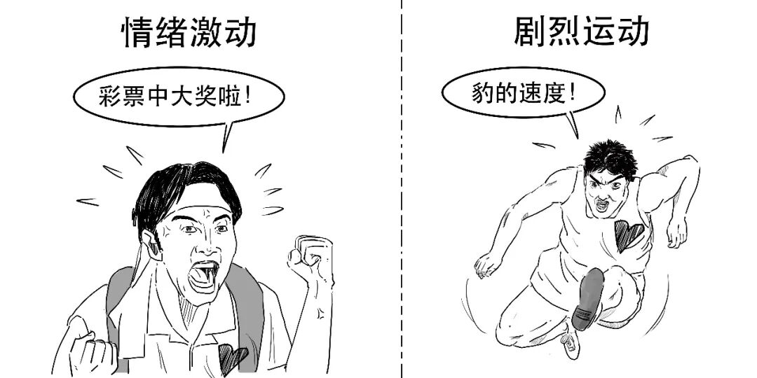
Sudden emotional excitement or strenuous exercise can raise pressure abruptly.
Now that we understand hypertension, where might a vessel rupture?
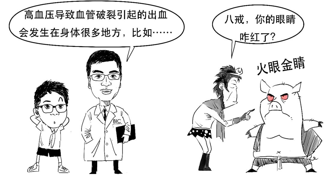
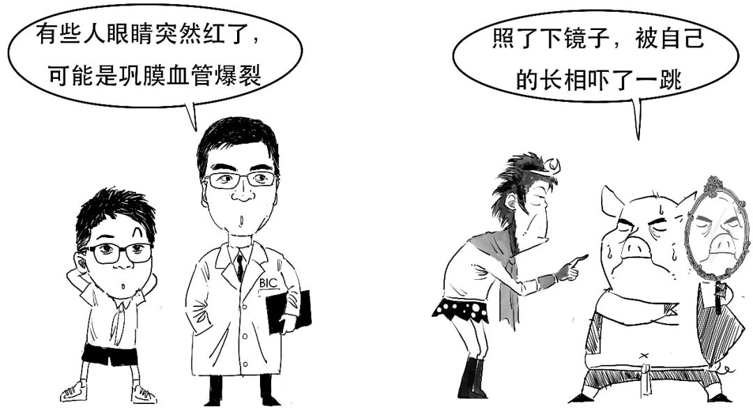
The sclera mentioned here is the white of the eye. We now focus on vessel rupture inside the head.
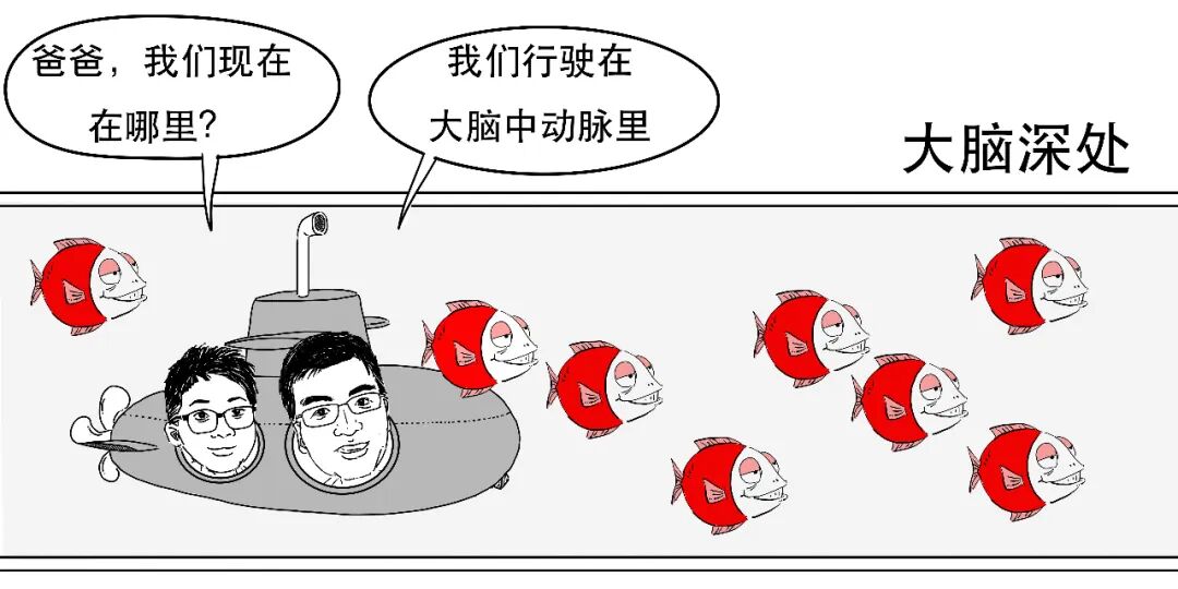
The lenticulostriate arteries in the basal ganglia have a distinctive course, almost perpendicular to their parent artery, an unusual arrangement among blood vessels.
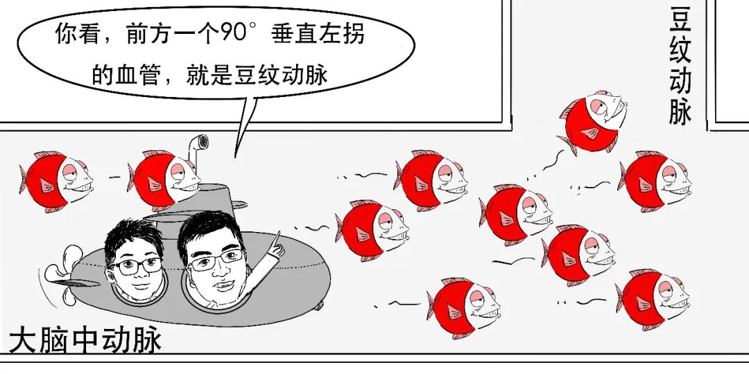
At this near-right-angle turn, each red blood cell is like a vehicle drifting around a bend. With hypertension, high-pressure flow strongly impacts this area.
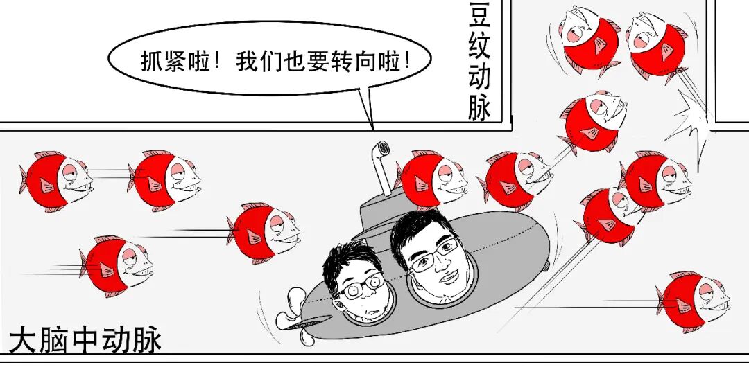
Over time, if the vessel cannot withstand pressure, it may rupture. The lenticulostriate arteries are in the basal ganglia, making this a common location for hypertensive brain haemorrhage.
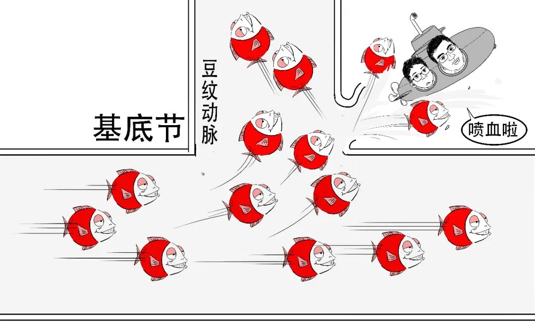
The basal ganglia region also lies along pathways transmitting signals from the cortex to the limbs.
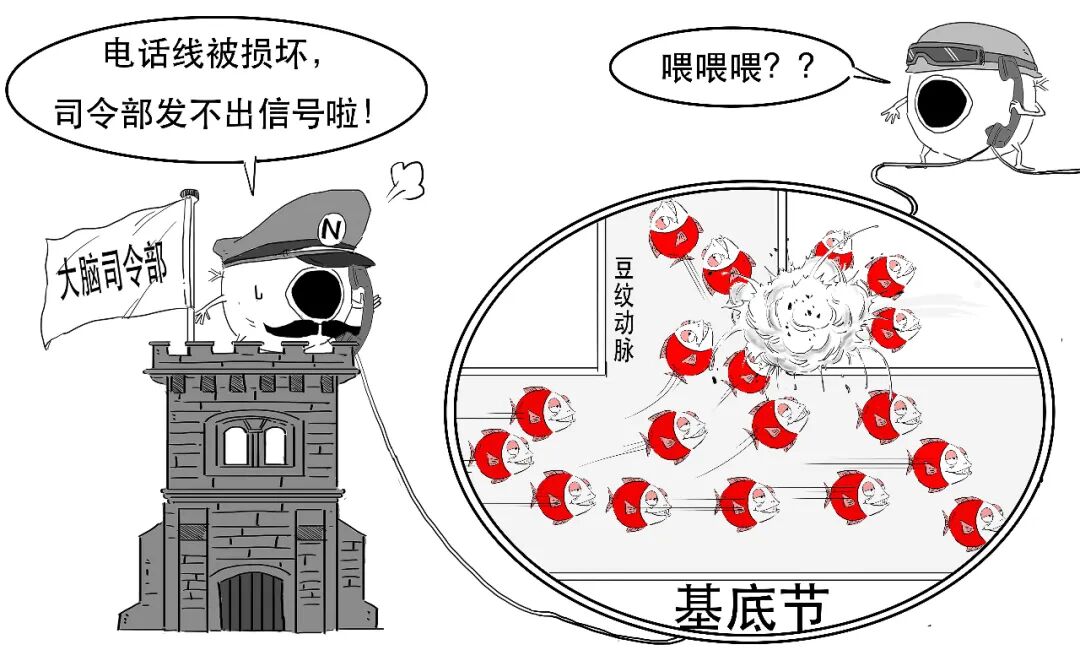
Bleeding can damage and block those pathways, preventing movement commands reaching the limbs. One side may become immobile. Hemiplegia or impaired movement on one side is therefore a common symptom.
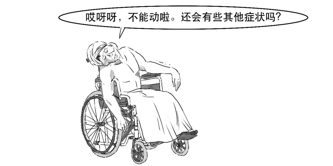
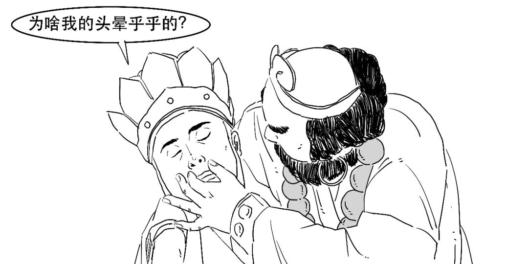
Tissue damage and haematoma compression may also cause severe headache, dizziness and projectile vomiting associated with increased intracranial pressure. Consciousness may change as well.
The ventricles containing cerebrospinal fluid are next to this region. Blood entering them may obstruct fluid circulation, cause acute hydrocephalus and impair consciousness. For our earlier discussion, see “Hydrocephalus Explained in Comics.”
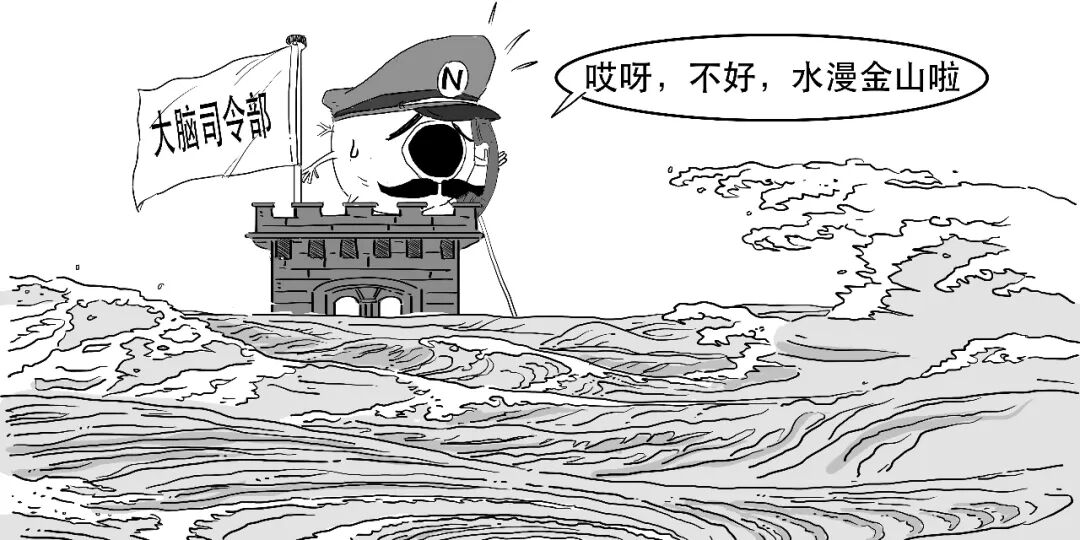
Impaired consciousness and a larger bleed require particular concern.
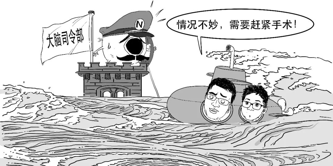
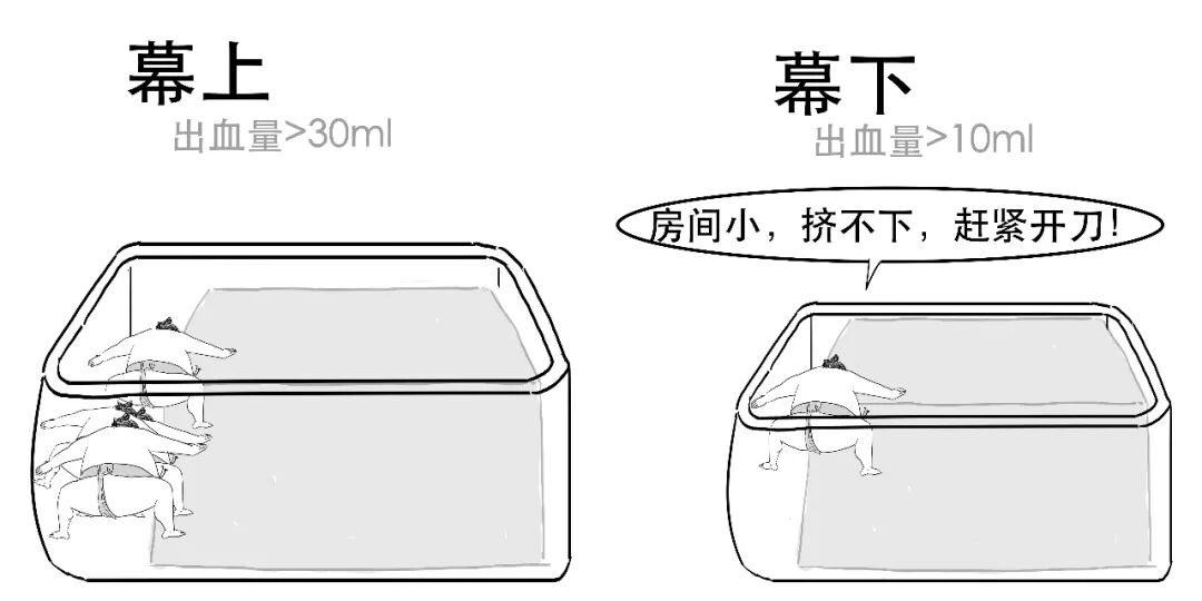
The source gives examples of considering surgery when supratentorial bleeding exceeds 30 mL or infratentorial bleeding exceeds 10 mL.
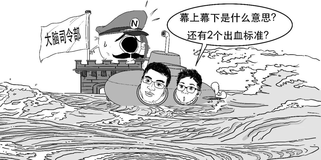
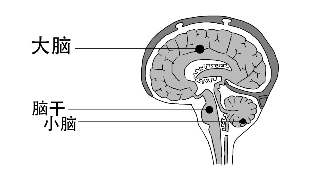
This involves an anatomical distinction. First, look at the head.
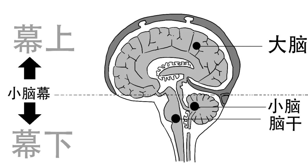
A tent-like partition separates the cerebrum and cerebellum.
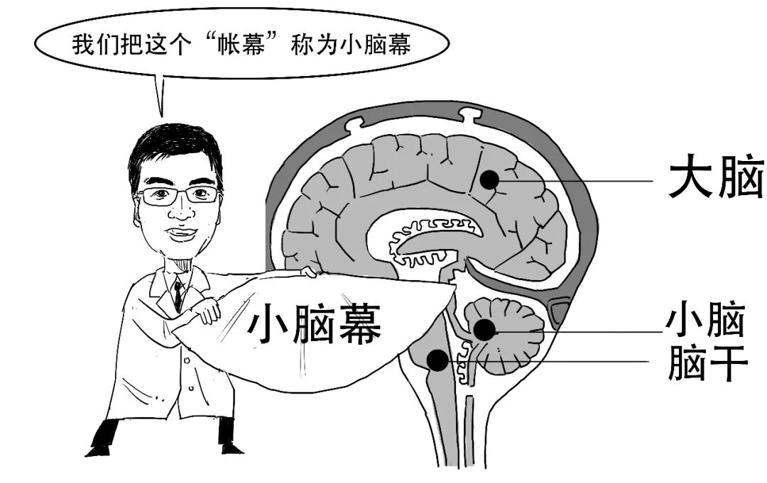
Above the tentorium is the supratentorial region, mainly the cerebrum. Below it is the infratentorial region, including cerebellum and brainstem.
The smaller infratentorial space means even a smaller haemorrhage may compress vital brainstem centres. The article therefore contrasts consideration of surgery above 10 mL there with above 30 mL supratentorially.
Surgery aims to stop haematoma progression through haemostasis and relieve compression through evacuation, allowing compressed tissue some relief.
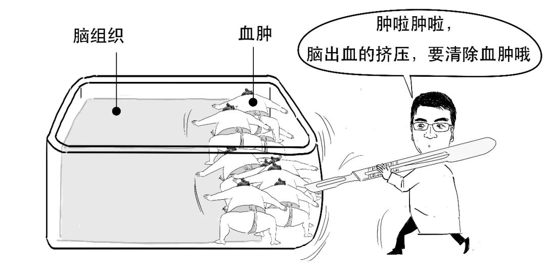
A large bleed may require craniotomy and haematoma removal; a smaller one may sometimes be drained through a tube. These concepts were discussed earlier in “Craniotomy Explained in Comics.”
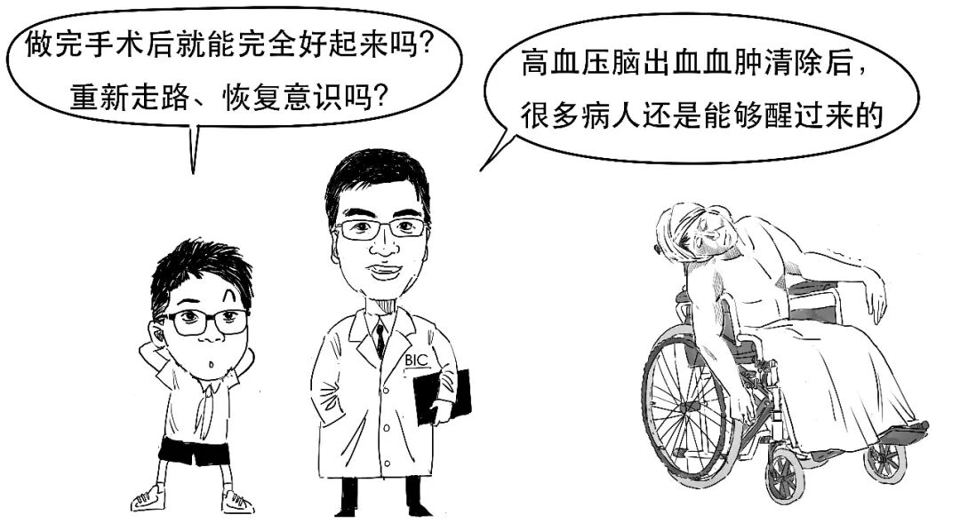
Recovery depends on many factors: volume and location, whether surgery is timely and well performed, postoperative critical care and rehabilitation.
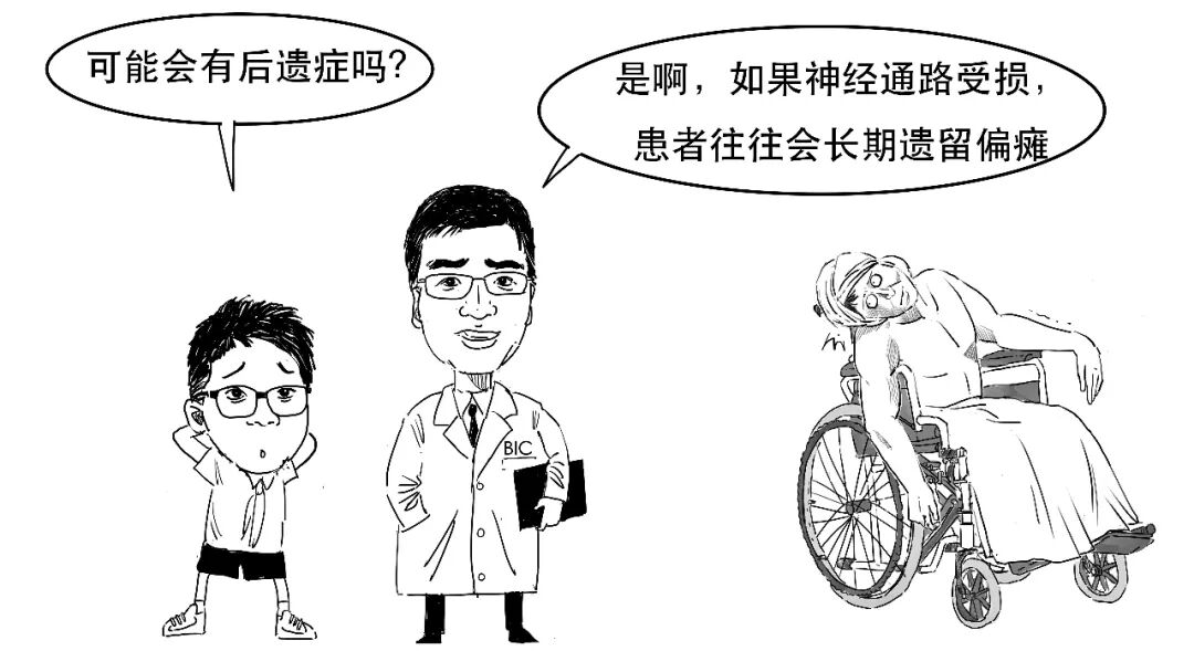
Sequelae include neurological impairment, especially movement and speech, and secondary hydrocephalus. Injury to the dominant hemisphere may leave language difficulties.
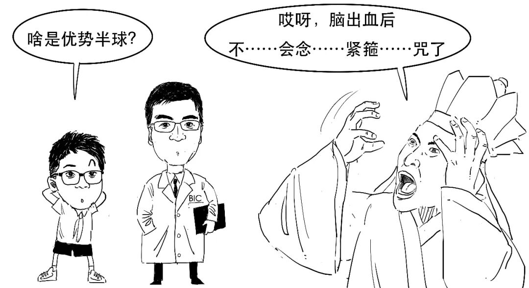
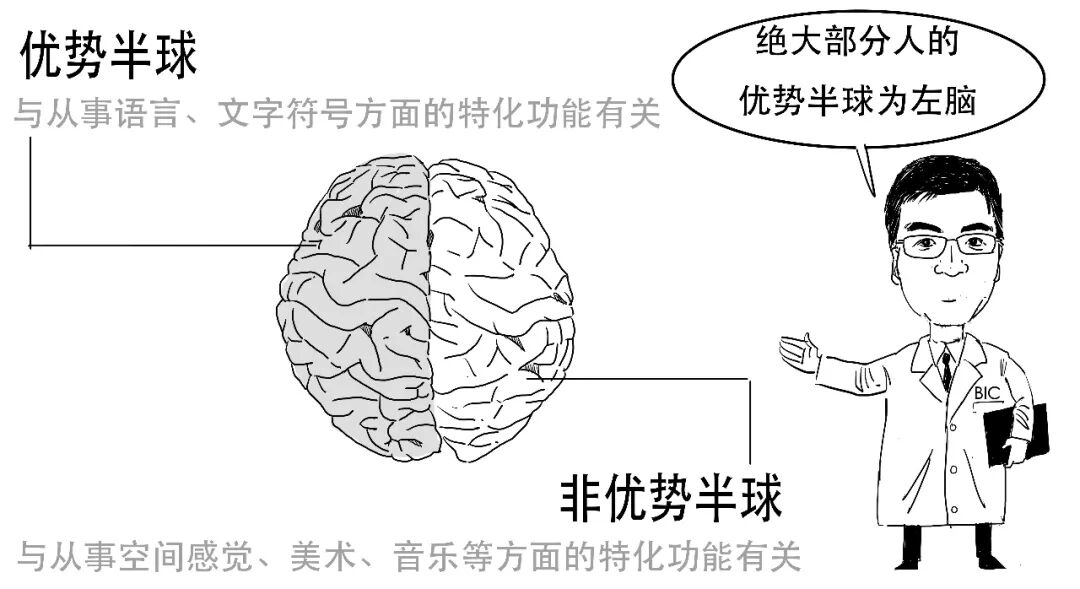
The brain has dominant and non-dominant hemispheres, and dominance differs between people.
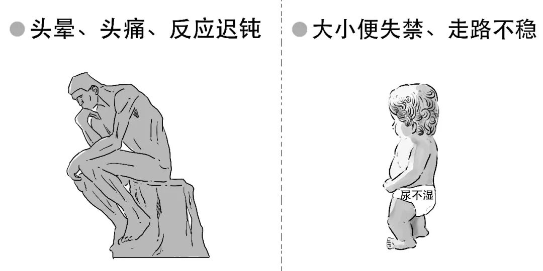
Even after surgery, bleeding can disrupt fluid circulation and the balance of cerebrospinal fluid production and absorption. The original article states that around 35% may develop secondary hydrocephalus, with principal symptoms illustrated below.
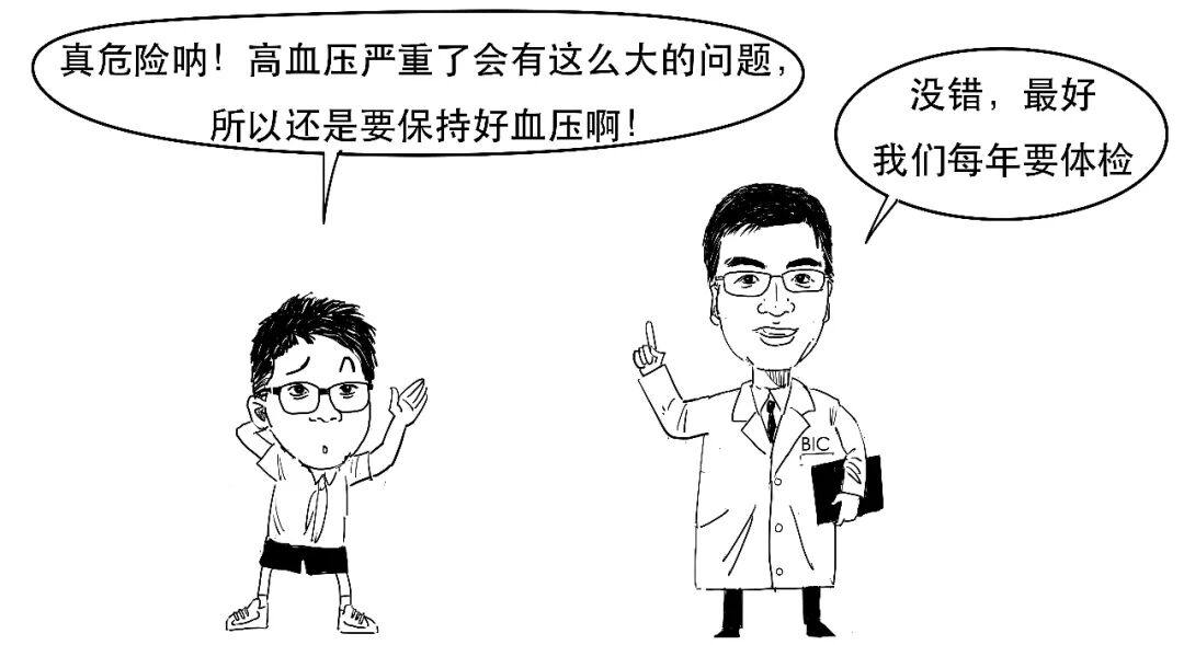
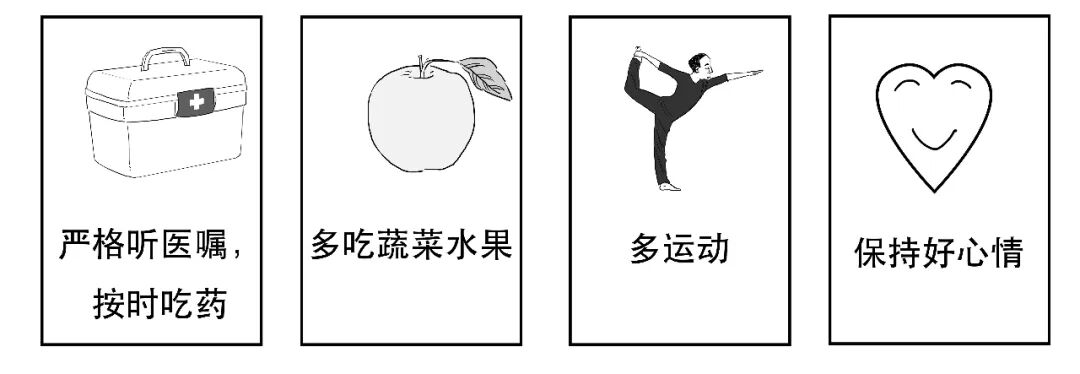
After a hypertension diagnosis, there is no need for excessive panic; the following illustrations explain actions to take.
Previous instalments
1. Traumatic Brain Injury in Comics. 2. Craniotomy in Comics. 3. Neuroscience Technology in Comics. 4. Hydrocephalus in Comics.
Disclaimer
This is a serial neuroscience education comic. We strive for accurate and reliable information, but it is not medical consultation advice and should not be used as a diagnostic standard for neurosurgical diseases. For related conditions, seek care at a qualified hospital.

高血压这三个字对我们来说并不陌生。
判定高血压要经过2次有间隔地测量,血压都高才算高血压。
我们今天就来唠一唠高血压,以及高血压导致的脑出血。
高血压作为一种疾病,是一种长期的状态。
高血压就类似水龙头一直开的很大,水管里的压力一直都很高。
人的血液在血管里流淌,就像水在水管里流动一样,会对管壁产生压力。
有一些人患有高血压,他们的血流动力会发生变化,血液对血管的压力就会比一般人要大,某些部位的血管逐渐变脆弱。
那到底多高的血压算高血压呢?
我们可以把血压分为收缩压和舒张压。
收缩压就是心脏在收缩的时候血液对血管壁的压力,而舒张压就是心脏在舒张的时候血液对血管壁的压力。
就像给气球打气一样,收缩压对血管壁的压力要大于舒张压。
高血压就是收缩压或者舒张压达到了一定大小,
标准如下:
对于高血压患者来说,
如果血管壁长期承受着高压的损伤,在某一天血压突然升高的时候,血管就可能会破裂出血。
当我们在情绪突然激动或者剧烈运动的时候,都会导致血压突然增高。
理解了高血压,
那你知道高血压一般会导致哪儿的血管破裂吗?
这里说的巩膜,就是白眼球部位。
下面我们重点说说高血压导致脑袋里血管破裂出血的情况。
大脑血管中有一个非常特殊的部位,叫基底节的豆纹动脉。
这个动脉很特别,是因为它的走向跟它起源的大动脉几乎是垂直的,这在人体所有血管中都是极其罕见的构造。
在这个豆纹动脉的垂直弯道,每个流过的红细胞都要像经历一个“漂移过弯”。
如果患有高血压,大量血流的压力就会强力冲击到这个特殊的垂直部位。
长此以往,一旦这里的血管承受不住压力,就会破裂出血。
而这里豆纹动脉所在的区域,正是基底节部位。
因此,基底节部位的脑出血也是最多发的高血压脑出血。
而基底节正好是大脑皮层支配肢体的神经向下延伸、传递神经电信号的必经之路。
这里出血损伤了,这条神经传导的路就被封了,给肢体发出指令的神经电信号就被拦在大脑里,没法继续传导给肢体。
这时候,患者身体的一侧就动不了了。
所以,偏瘫或一侧肢体运动功能障碍是高血压脑出血的常见症状之一。
然而,脑出血后由于脑组织的破坏、血肿的压迫,
常会伴有剧烈的头痛、头晕、喷射样呕吐等颅内压增高的表现。
同时,还可能会有意识障碍变化,
这是因为基底节部位旁边就是我们脑脊液所在的脑室系统,出血一旦破入脑室,阻碍了脑脊液的循环,会引发所谓的急性脑积水,从而造成意识障碍。
关于脑积水我们以前说过,想了解的同学点这里:
一般情况下,如果高血压脑出血的患者存在意识障碍、出血量也比较多,就要格外小心,
比如,幕上出血量大于30ml、幕下出血量大于10ml,我们要开始考虑去做手术了。
这就涉及到解剖学的一个概念。
首先来看下我们脑袋的样子:
在大脑与小脑之间有一层间隔,形状类似帐幕,
在小脑幕上方的,我们称为幕上,主要是大脑;
而小脑幕下方的,我们称为幕下,包括小脑、脑干。
相对于幕上,幕下的空间更小,稍微出点血,就会压迫脑干的生命中枢,所以幕下的出血量大于10ml就要考虑做手术了;而一般幕上出血量大于30ml才会考虑。
高血压脑出血手术治疗的目的是解除血肿的进展(止血)和压迫(清血肿),让患者被压迫的脑组织喘口气,得到一定的缓解。
如果出血量大,甚至要“开大刀”,要把骨头拿掉,进行开颅血肿清除术。出血量小,也可以“开小刀“,比如用一根管子把血抽出来。
这部分内容在以前章节讲过,想看的点下面链接,这里就不展开了。
但恢复的好坏也要考虑许多因素。
比如,出血量多少、出血部位、手术是不是及时做、手术是不是做得好、手术后的重症管理是不是到位,以及康复锻炼,对后面恢复情况都很关键。
高血压脑出血的后遗症包括神经功能(主要是运动、言语)障碍和继发性脑积水。
比如,如果脑出血损伤到了优势半球,可能会遗留语言功能障碍。
我们把大脑分为2个半球,分别是优势半球和非优势半球,不同的人优势半球还不一样。
又比如,即使动了手术,但由于脑出血可能影响正常脑脊液循环,打破脑脊液的生成-吸收平衡,有35%左右的患者可能会出现继发性脑积水,
主要症状包括:
如果一旦确诊高血压,
我们也不必过分惊慌,需要做好以下动作:
往期精彩内容回顾:
1、“漫”谈脑外伤
2、“漫”话开颅术
3、“漫”演神科技
4、“漫”引脑积水
免责声明
本文为神经科学科普连载漫画,我们力求相关信息准确可靠,但不将此科普内容作为求医问诊的建议,读者也不应以此漫画作为神经外科疾病判断的标准。如有相关疾病,请至正规医院就诊。