Hydrocephalus explained: cerebrospinal fluid and shunt surgery
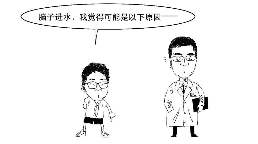

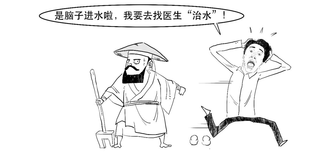
People often jokingly say someone has “water in their brain”. Can the brain really fill with water?
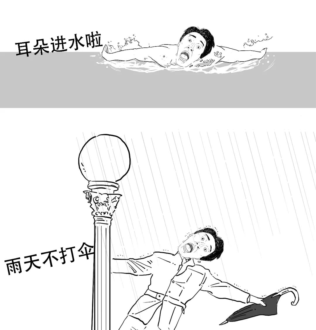
There really is “water” inside the brain, but it has not flowed in from outside!
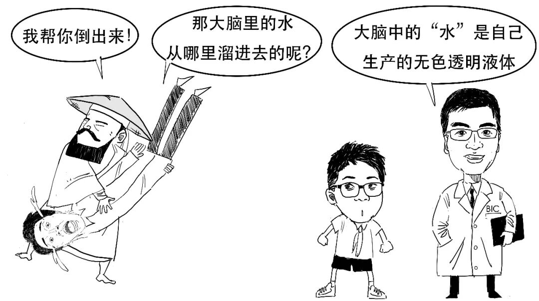
This fluid is called cerebrospinal fluid, or CSF.
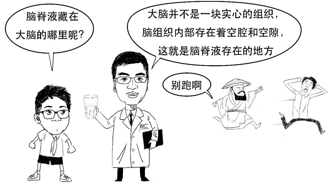
CSF is found in three places in the body.
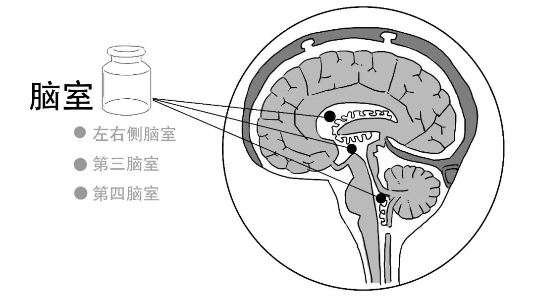
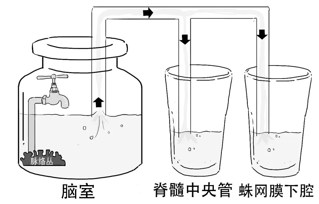
1. The ventricles.
These cavities in the centre of the brain are filled with CSF.
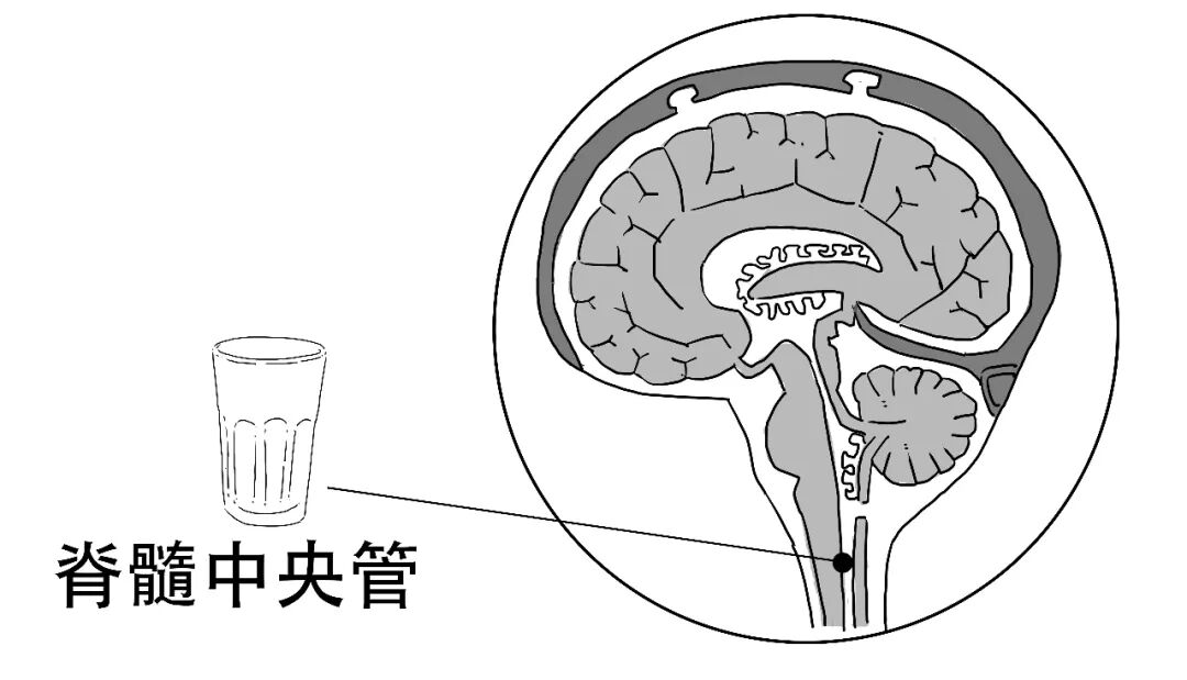
2. The spinal cord's central canal.
The central nervous system comprises the brain and spinal cord. The cavity inside the spinal cord's central canal also contains CSF.
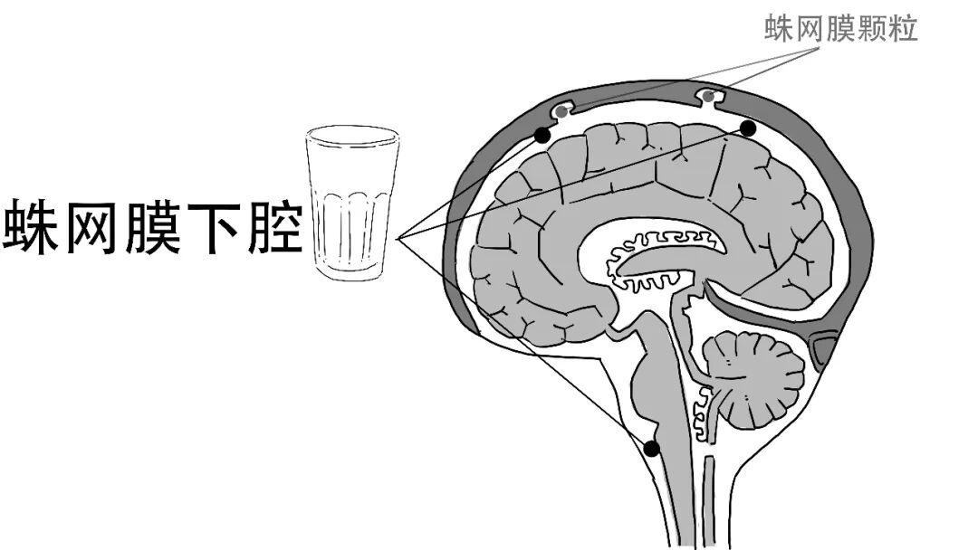
3. The subarachnoid space.
The space beneath the arachnoid membrane also contains CSF.
CSF protects the soft central nervous system, cushioning external impacts that could injure the brain and spinal cord. It is a little like this:
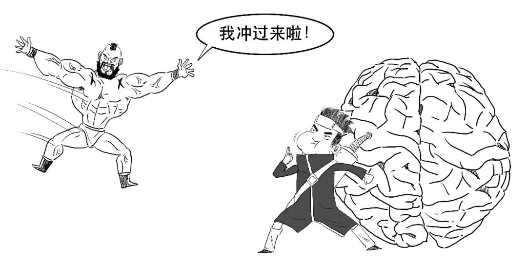
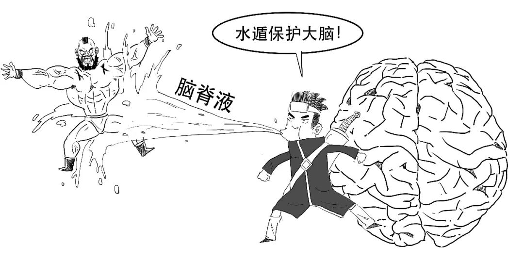
Together, CSF in these three locations protects the brain and spinal cord, rather like a baby surrounded by amniotic fluid.
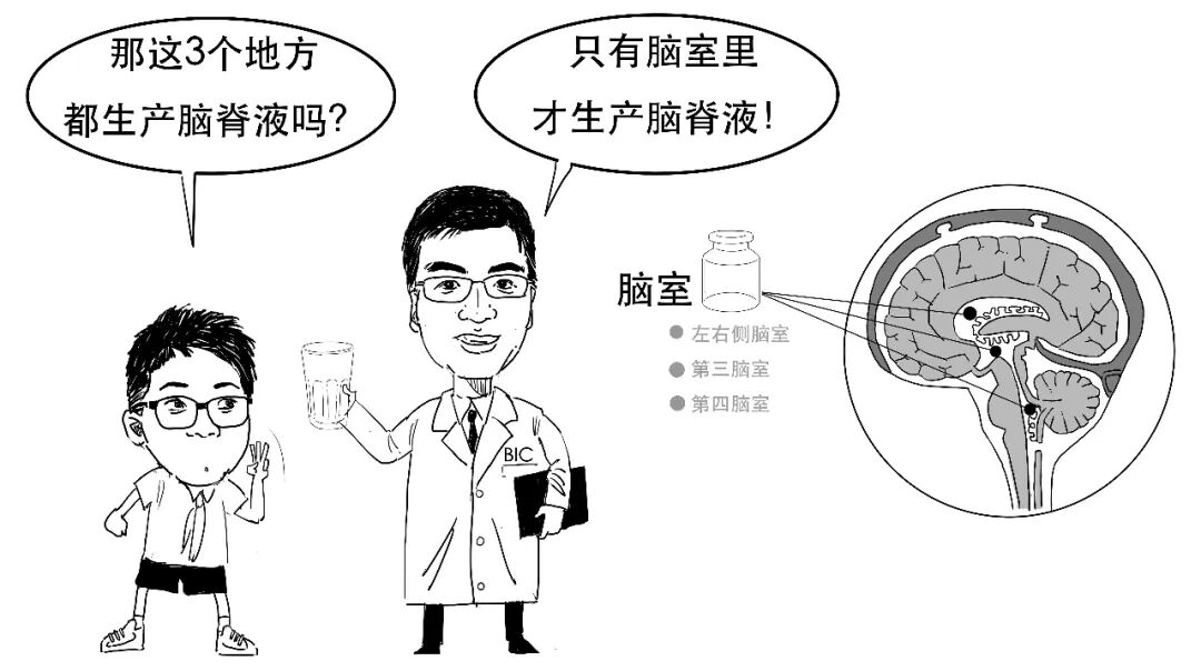
CSF is produced by the choroid plexus in the ventricles. The original comic describes the choroid plexus as a venous structure in the brain and explains CSF as a body fluid derived from venous blood.
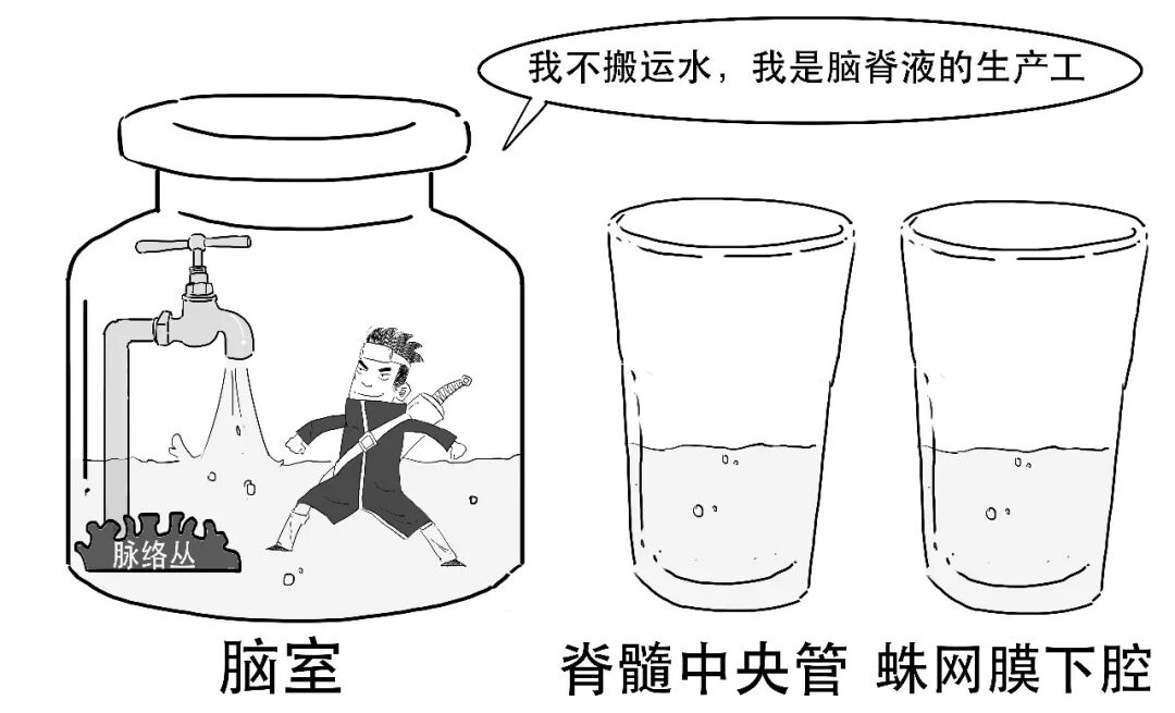
The “water” produced in the ventricles flows to the other two locations, covering the spinal cord and brain surface.
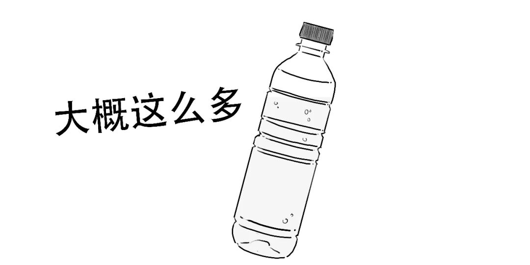
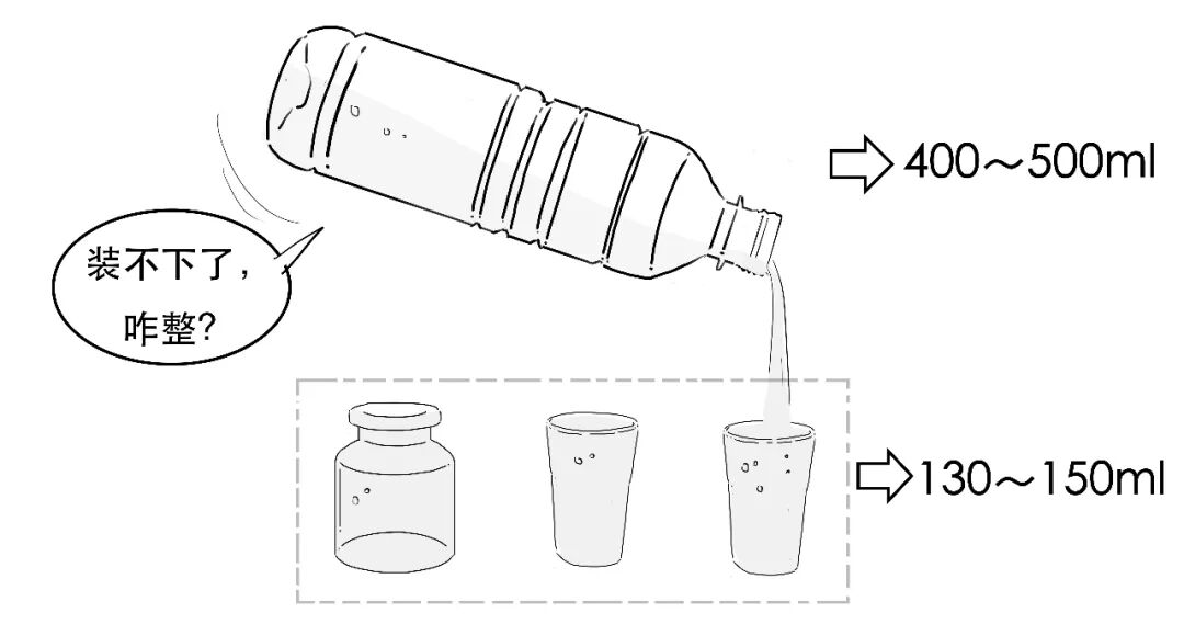
An adult can produce 400–500 mL of CSF each day.
Yet an adult has room for only 130–150 mL of CSF.
After entering the subarachnoid space, CSF can be absorbed through arachnoid granulations and returned to venous blood.
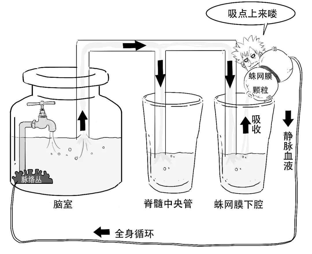
Through circulation and recycling, CSF washes away metabolic waste from nervous tissue, much as blood does.
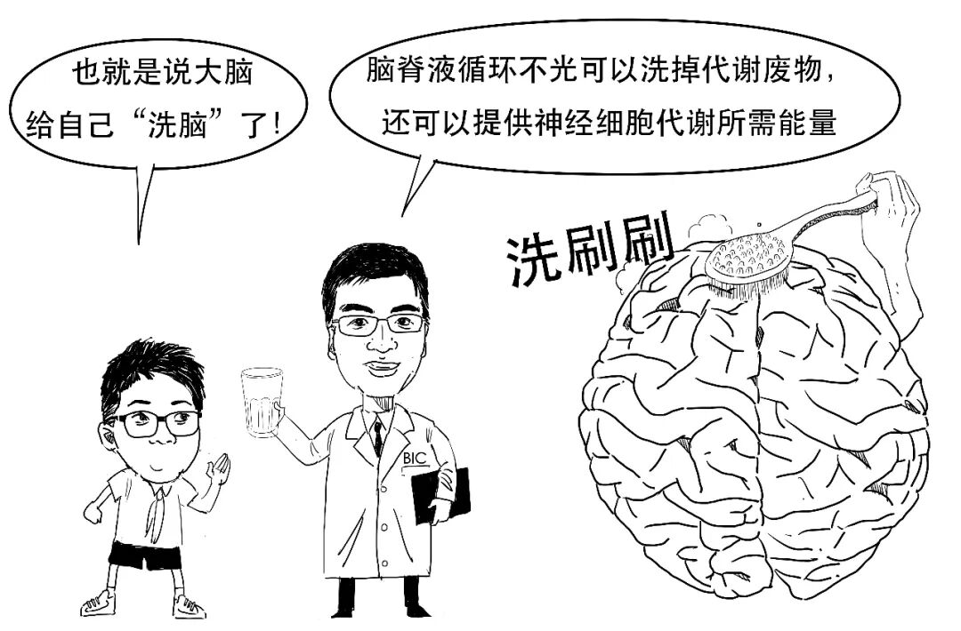
CSF is therefore not only the brain's protector and cleaner, but also its energy station!
If the brain needs this “water”, why do doctors need to control it?
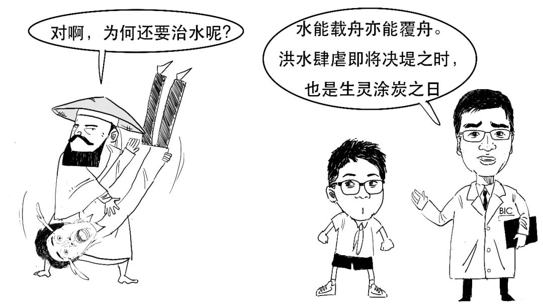
Too much CSF causes hydrocephalus, expanding the ventricles, compressing brain tissue and harming the nervous system.
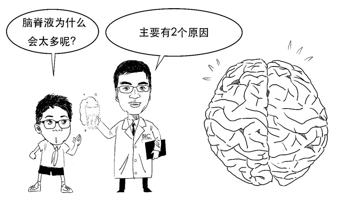
1. CSF cannot leave the ventricles.
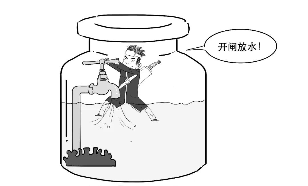
Fluid accumulates inside a ventricle because it cannot flow out. This is called obstructive hydrocephalus.
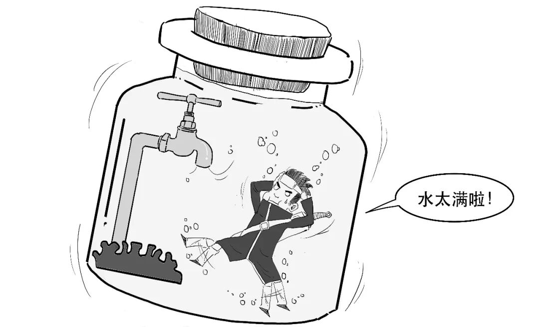
2. Too little CSF is absorbed.
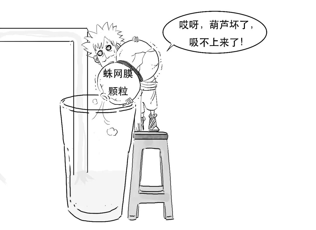
Fluid leaves the ventricles, but damaged arachnoid granulations cannot absorb it during circulation. This is called communicating hydrocephalus.
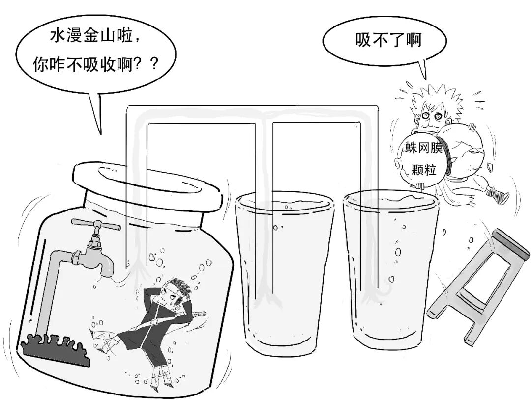
CSF production and absorption normally balance one another. Disrupting that balance can cause excessive fluid and hydrocephalus.
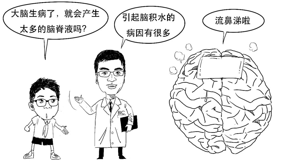
Causes may include congenital conditions, TBI, brain haemorrhage, brain tumours and intracranial infection. Some cases have no clear cause and may be related to autoimmune responses.
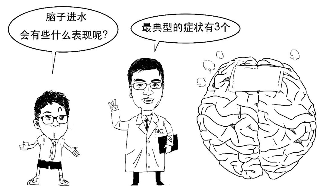
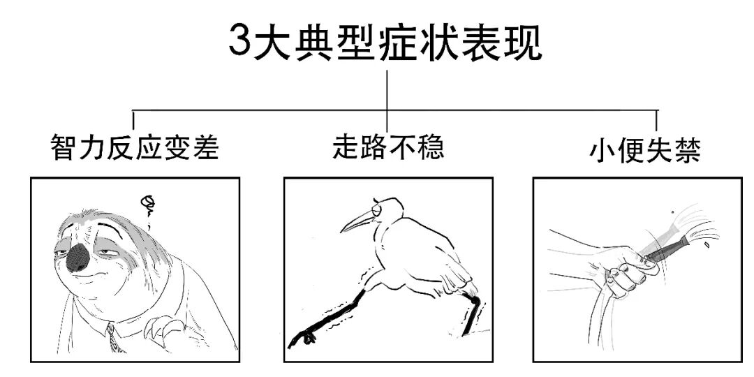
When patients develop these symptoms, neurosurgeons consider whether hydrocephalus is present.
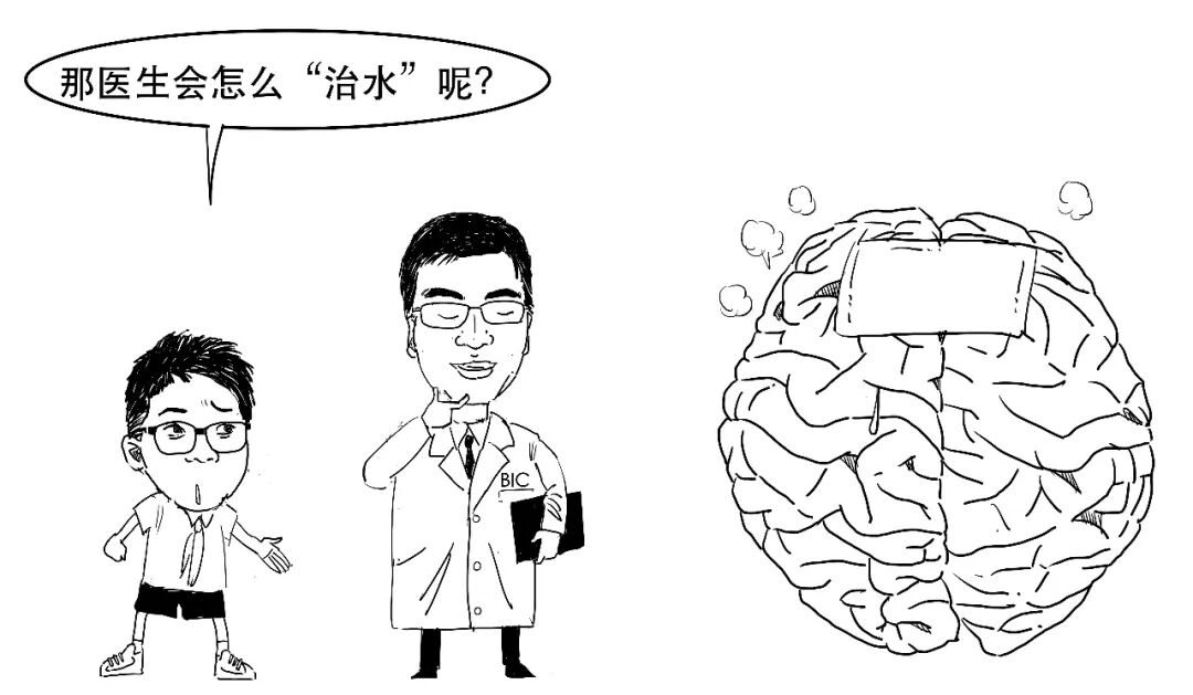
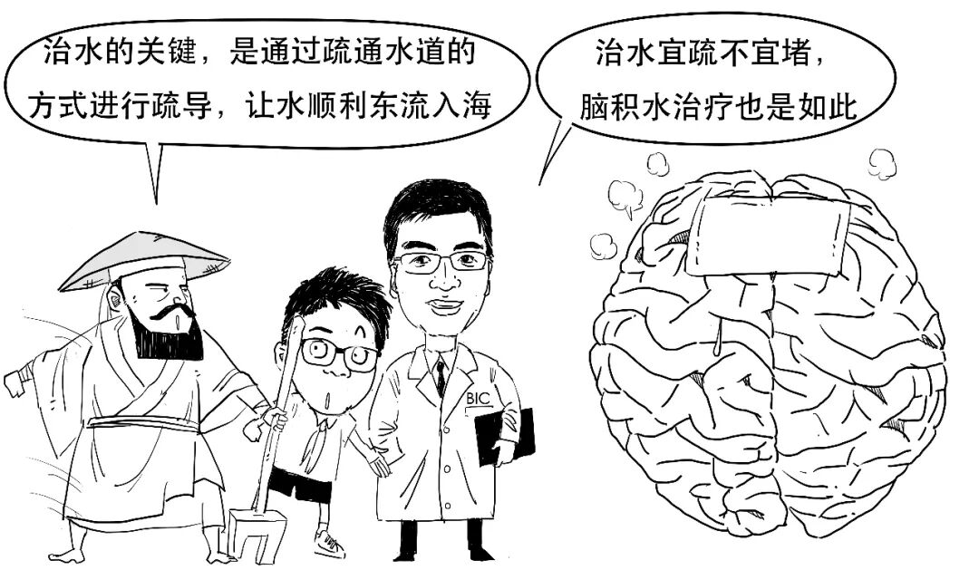
The main treatment is surgery to shunt excess CSF outside the ventricles. The most common shunt procedure places a catheter into a ventricle,
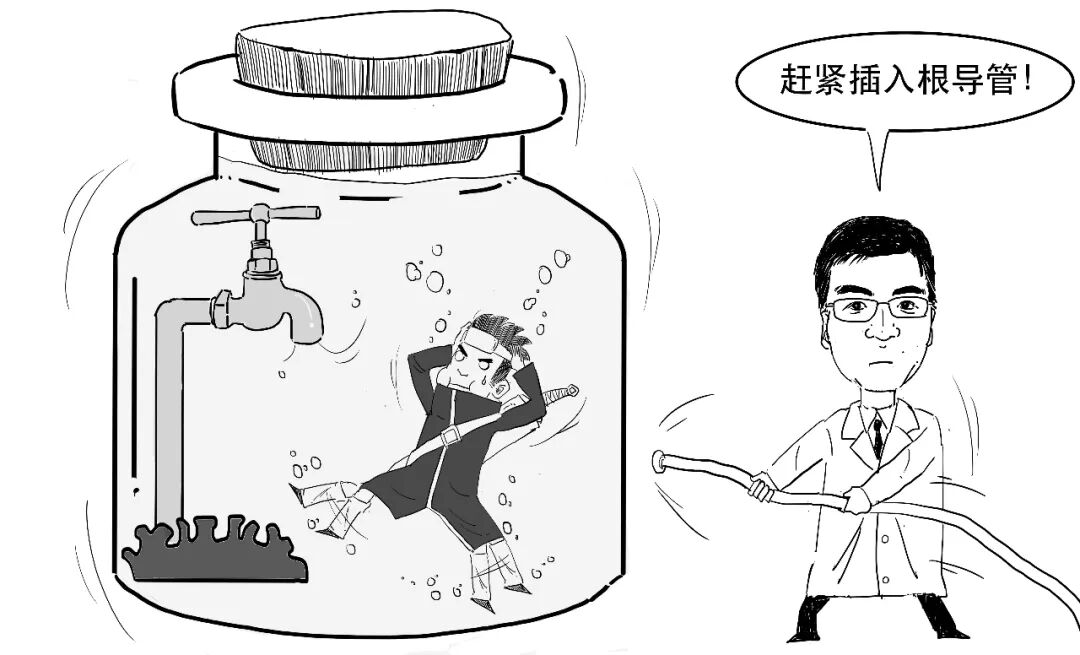
then drains the excess fluid through it to another part of the body, such as the abdomen or an atrium of the heart.
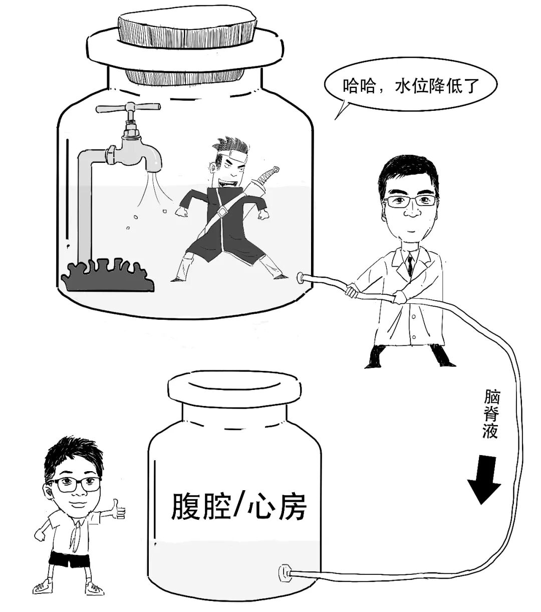
CSF shunted to the abdominal cavity can be absorbed there or enter the circulation.
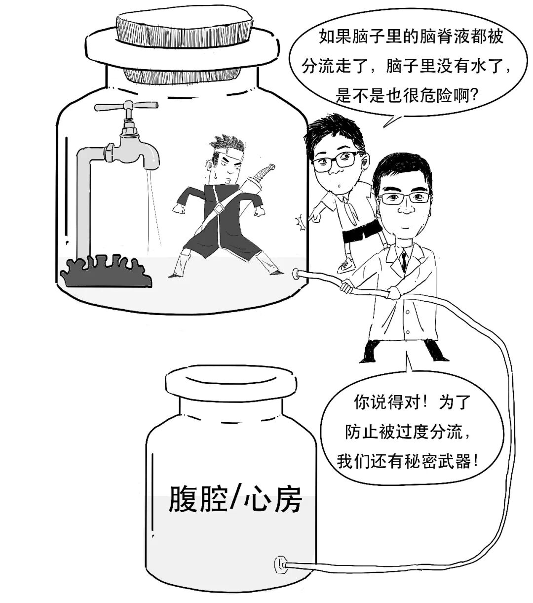
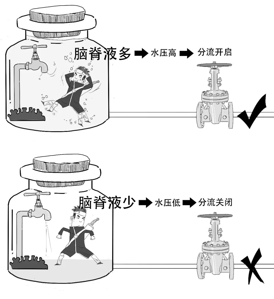
A valve on the shunt catheter senses pressure and regulates drainage.
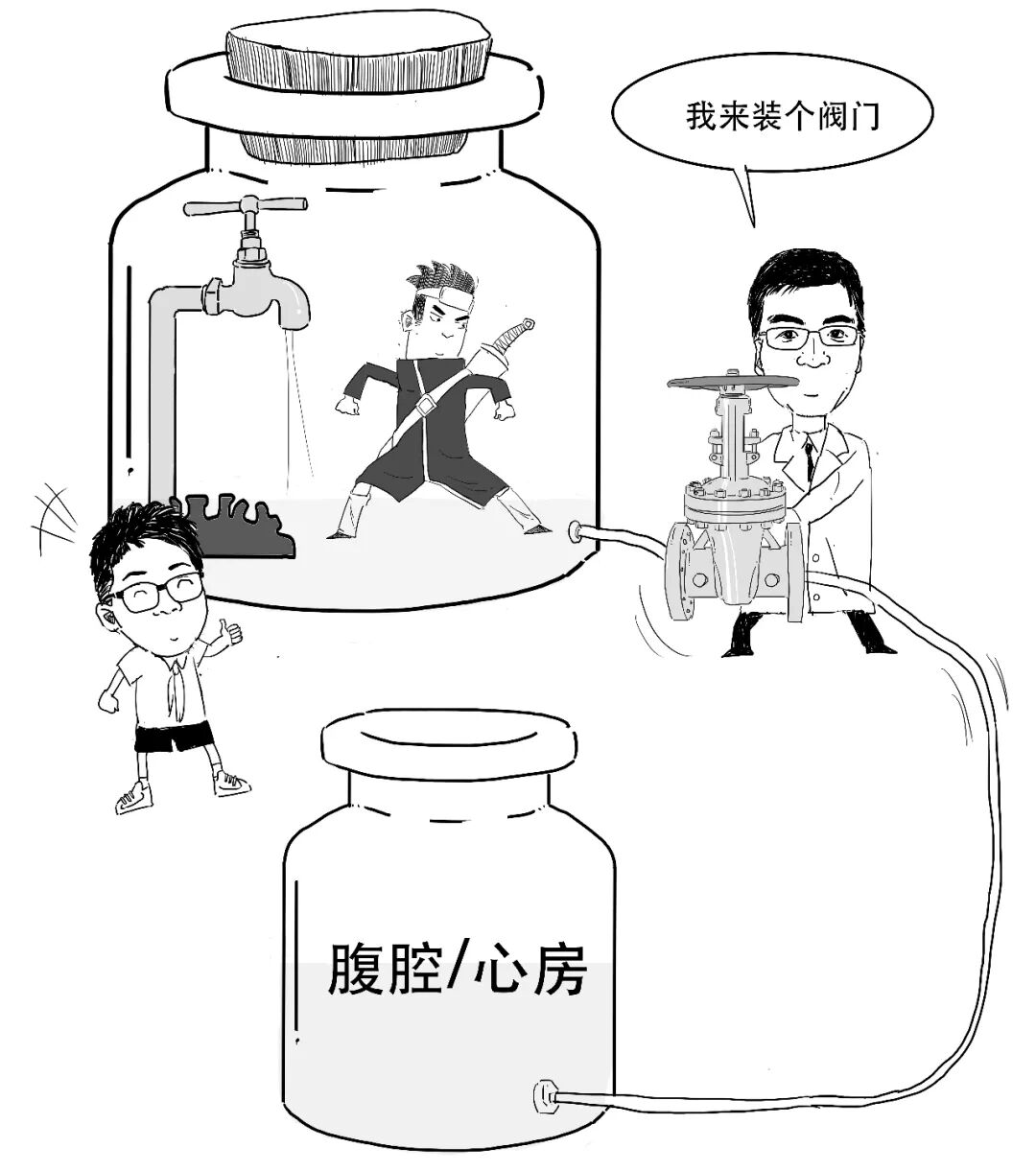
As CSF volume decreases, its pressure falls. When pressure drops below the valve threshold, the shunt closes, helping maintain a normal amount of fluid in the brain.
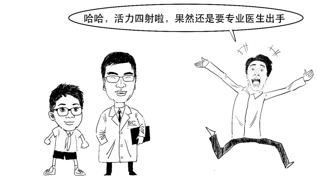
Once the accumulated fluid has been relieved in this way, the patient can gradually recover.
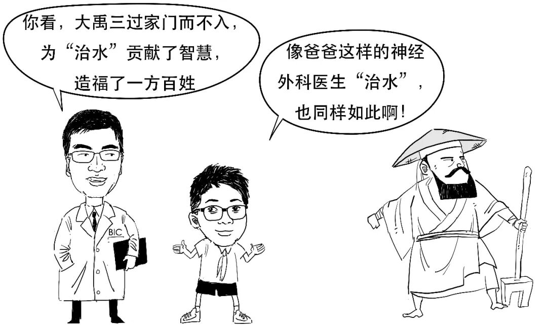
Although shunt surgery is relatively minimally invasive, intracranial infection or shunt obstruction can still occur afterwards and may require another neurosurgical procedure.
Previous comic instalments: “Talking About Brain Injury”, “Understanding Craniotomy” and “Neuroscience and Technology in Comics”.
Disclaimer
This is a serial neuroscience education comic. We aim for accurate, reliable information, but it is not advice for seeking medical consultation, and readers should not use it to judge neurosurgical disease. If you have a related condition, attend a qualified hospital.
我们总听人说脑子进水啦,
但脑子真的会进水吗?
大脑里确实有“水”,
但可不是外面流进去的哦!
我们把这种液体
叫做脑脊液(cerebrospinal fluid,CSF)。
人体有3个地方可以有脑脊液,分别是
/1/ 脑室
脑室位于大脑的中央位置,里面是空腔,充满着脑脊液。
/2/ 脊髓的中央管
人的中枢神经系统包括脑和脊髓。
在脊髓中央管内的空腔中同样也有脑脊液。
/3/ 蛛网膜下腔
蛛网膜的下腔有空隙,里面也有脑脊液。
脑脊液可以保护柔软的中枢神经,缓冲外界冲击对脑和脊髓造成的损伤。
这种感觉有点像:
因此,这3个地方的脑脊液就让整个脑、脊髓组织像是泡在羊水里的婴儿一样,受到全面保护。
脑脊液是通过脑室中的脉络丛结构来生成的。
脉络丛结构是一种脑内静脉结构。因此,脑脊液其实是一种来源于静脉血液的体液。
脑室里生产出来的“水”,再流到了另外2个地方,从而使脑脊液覆盖到整个脊髓和脑表面。
成人每天可以产生400~500毫升的脑脊液。
但是,成人仅有130~150毫升容纳脑脊液的空间。
其实,脑脊液进入到蛛网膜下腔之后,脑脊液可以被蛛网膜上的蛛网膜颗粒吸收,然后再回到静脉血液中。
经过这样的循环和回收,脑脊液像血液一样冲洗带走了神经组织的代谢废物。
所以说,脑脊液不光是大脑的守护者、清洁工,还是大脑的能源站呢!
既然大脑的运行离不开这种“水”,那医生为何还要去“治水”呢?
如果脑脊液过多,就会引发“脑积水”,把脑室结构撑大,挤压脑组织,会对神经系统有损害。
/1/ 脑脊液无法流出脑室
这时候,水积在脑室内部,堵在里面流不出去,这种就被称为“梗阻性脑积水”;
/2/ 脑脊液吸收太少
这时候,水虽然流出了脑室,但在循环过程中,负责吸收脑脊液的蛛网膜颗粒损坏了,无法吸收脑脊液,这种被称为“交通性脑积水”。
脑脊液生成和吸收存在着一种平衡关系,这种平衡被打破就会引起脑脊液过多,导致脑积水。
比如有先天性、脑外伤、脑出血、脑肿瘤、颅内感染等原因,或者是不明原因发生的,可能与自身免疫反应相关。
如果病人出现这些症状,
神经外科医生就会考虑有没有脑积水了。
治疗脑积水最主要的手段,就是通过手术的方式将多余的脑脊液“分流”至脑室外部。
最常见的分流手术方式是通过植入一根导管到脑室中,
然后通过导管将多余的脑脊液引流到身体内其他部位,包括腹腔、心房等。
假如脑脊液被分流进入腹腔,就可以被腹腔吸收或进入血液循环。
医生会在分流的导管上会安置一个阀门装置,通过感受压力来调节分流。
当脑脊液的容量减少后,相应的水压(脑脊液压力)就会下降。当压力下降至阀门阈值以下,脑脊液分流关闭,就能维持脑内正常的脑脊液。
通过上述方式,
等到脑积水的“水”疏通之后,病人就能慢慢康复了。
尽管分流手术创伤较小,但手术完成后依然有可能出现颅内感染、分流堵塞等并发症,此时必须要神经外科医生再次手术处理。
往期精彩内容回顾:
1、“漫”谈脑外伤
2、“漫”话开颅术
3、“漫”演神科技
免责声明
本文为神经科学科普连载漫画,我们力求相关信息准确可靠,但不将此科普内容作为求医问诊的建议,读者也不应以此漫画作为神经外科疾病判断的标准。如有相关疾病,请至正规医院就诊。
欢迎点击“”。